Our Research
Iron is an essential metallonutrient for nearly all living organisms and represents a primary driver of host-pathogen interactions. During infection, pathogenic bacteria must be able to manage their requirement for iron with its potential for toxicity. This is achieved through a combination of iron uptake and regulatory pathways, which function to maintain iron homeostasis. The focus of our laboratory’s research is to understand how these systems mediate iron homeostasis and contribute to bacterial pathogenesis. Our studies are currently focused on Pseudomonas aeruginosa and Staphylococcus aureus, opportunistic pathogens of significant concern for hospital-acquired infections and in cystic fibrosis patients. We use a multidisciplinary approach to determine how iron homeostasis is mediated by these pathogens, as well as the impact of these systems on pathogenesis.
Pseudomonas PrrF sRNAs

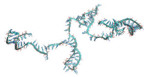
A long-standing interest of my laboratory is understanding how small regulatory RNAs (sRNAs) contribute to iron homeostasis of P. aeruginosa. P. aeruginosa transcribes two iron-responsive sRNAs, named PrrF1 and PrrF2, which block the expression of iron-containing proteins in iron-depleted environments. In doing so, the PrrF sRNAs “budget” the use of this essential nutrient when it becomes scarce. We have also demonstrated that the PrrF sRNAs allow for the production of the Pseudomonas quinolone signal (PQS), a quorum sensing molecule required for activation of multiple virulence genes. Thus, the PrrF sRNAs exert broad effects on cell physiology and virulence traits.
The PrrF sRNAs are distinct from many iron-responsive bacterial sRNAs in that the two nearly identical PrrF RNAs are transcribed from genes located in tandem on the P. aeruginosa chromosome. This arrangement allows for the expression of an additional, longer RNA, designated PrrH, which is responsive to heme as well as iron. Since heme constitutes a significant proportion of iron during infection, we hypothesize that the PrrH sRNA imparts unique regulatory functions that are important for pathogenesis. We are currently working to understand 1) how the PrrH sRNA is expressed and regulated by heme, and 2) how PrrH regulation contributes to P. aeruginosa physiology and virulence.
Iron Homeostasis in the CF Lung

P. aeruginosa is responsible for life-long and debilitating chronic lung infections in cystic fibrosis (CF) patients. Within the CF lung, P. aeruginosa evolves considerably and becomes increasingly resistant to antibiotic therapy. P. aeruginosa uses a variety of strategies to acquire iron, one of which relies on a small iron-scavenging molecule called pyoverdine. Pyoverdine is required for acute infections, but the ability of some P. aeruginosa strains to make pyoverdine is lost after several years of chronic CF lung infection. In collaboration with the laboratory of Dr. Angela Wilks at UMB, we previously showed that heme acquisition is enhanced in CF isolates over time, indicating heme is a more readily available source of iron in the CF lung. We are currently working to understand the molecular basis of these adaptations.
Infection of the CF lung is not limited to P. aeruginosa, and several reports have demonstrated that interactions between distinct microbial pathogens affect the outcome of disease. In particular, the Gram-positive bacterium Staphylococcus aureus commonly infects younger CF patients. P. aeruginosa has been shown to lyse and use S. aureus as an iron source, and this process is dependent upon the genes that initiate synthesis of PQS. Our own studies have shown that this activity is enhanced in iron-depleted environments, indicating this “iron piracy” system is responsive to iron availability. In an effort to define the molecular basis of this phenomenon, we are collaborating with Dr. Maureen Kane, Director of the School’s Mass Spectrometry Center, to determine the impact of iron on the production of metabolites with both signaling and antimicrobial capabilities, combined with genetic analysis of iron regulatory pathways. These studies are expected to identify novel metabolites that drive polymicrobial interactions, as well as define the biosynthetic and regulatory pathways that mediate their production.
Recent collaborative studies with Dr. Elizabeth Nolan’s laboratory at MIT have also examined the impact of calprotectin (CP) on P. aeruginosa iron homeostasis. Originally identified as the “CF antigen” due to its abundance in the lungs of CF patients, CP makes up >40% of the soluble protein in circulating neutrophils. When released from neutrophils, CP binds to several divalent transition metal ions, including zinc, manganese, and nickel to hinder microbial growth. In 2015, the Nolan laboratory made the landmark discovery that CP also bound to the reduced, ferrous form of iron. Subsequent work in collaboration with our laboratory demonstrated that CP induces a robust iron starvation response in P. aeruginosa. Collaborative work in the Nolan and Oglesby laboratories, funded by NIH-NIGMS, aims to determine how CP-dependent sequestration of multiple metal ions, including iron, affects the physiology and virulence trait expression by P. aeruginosa.
